Tendon Diagram Foot - Anatomy Of The Foot And Ankle Orthopaedia - Overload heel pain, calcaneal fracture.
bymapatbrinson-
0
Tendon Diagram Foot - Anatomy Of The Foot And Ankle Orthopaedia - Overload heel pain, calcaneal fracture.. A tendon connects muscle to bone. They are often associated with activities requiring increased pressure on the ball of the foot, such as running, basketball, football, golf, tennis and ballet. Overload heel pain, calcaneal fracture. These tendons help your extensor muscles pull your foot upwards, which is necessary for walking. Foot tendinitis is a very common cause of pain due to irritation or inflammation in tendons of the foot after overuse or injury.
Exercises helps to improve its strength and work on the issues caused due to muscle tightness, shortening, or improper healing of a ligament. Sesamoid injuries can involve the bones, tendons and/or surrounding tissue in the joint. The ankle serves as foundation, shock absorber and propulsion engine. The muscles at the top of the foot fan out to supply the individual toes. The two main extensor foot tendons are the extensor hallucis longus and the extensor digitorum longus.
Foot Ankle Anatomy Pictures Function Treatment Sprain Pain from www.healthpages.org In addition, people with high arches are at risk for developing sesamoid problems. These tendons help your extensor muscles pull your foot upwards, which is necessary for walking. Ligaments are strong connective tissue composed of fibrous tissues. Torn ligaments can occur following a range of physical activities from dancing to snowboarding, and several common symptoms can help identify a torn ligament as the cause of your foot pain. Familiarity with the normal anatomy of the plantar tendons and its appearance at magnetic resonance (mr) imaging and ultrasonography (us) is essential for recognizing plantar tendon disorders. Tendinitis is treated with stretching and relaxing activities and exercises to give quality to the calf and foot muscles. The muscles at the top of the foot fan out to supply the individual toes. Muscles, tendons, and ligaments run along the surfaces of the feet, allowing the complex movements needed for motion and balance.
Foot tendinitis is a very common cause of pain due to irritation or inflammation in tendons of the foot after overuse or injury.
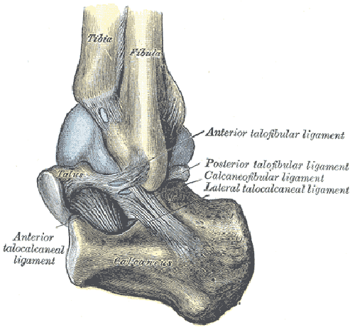
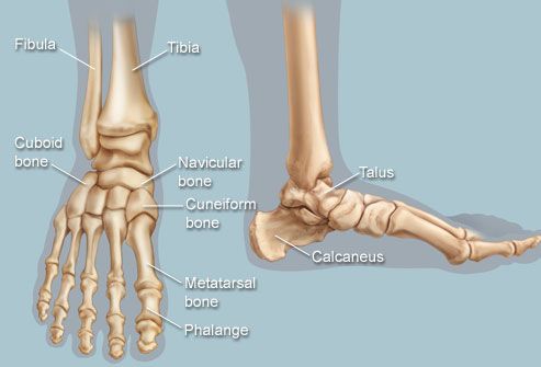
A tendon is a band of tissue that connects a muscle to a bone. A tendon connects muscle to bone. The human foot combines mechanical complexity and structural strength. Overload heel pain, calcaneal fracture. Ligaments stabilizing the ankle joint. Understanding the structure of the foot is best done by looking at a foot diagram where the anatomy has been labeled. Here you can see the tendons that extend down the top of your foot toward your toes, allowing you to curl your toes upward if need be. Other important tendons in the foot include the tibialis posterior (posterior tibial tendon), which attaches the calf muscle to the bones on the inside of the foot and supports the arch of the foot, and the tibialis anterior (anterior tibial tendon), which runs from the outer tibia to the first metatarsal and surfaces of the median cuneiform tarsal, which allows for dorsiflexion—bringing the. A tendon is band of tissue made up of many fibers. Allows the action of raising the foot. The calcaneus (heel bone) is the largest bone in the foot. Common ossicles of the foot some feet contain accessory ossicles or accessory bones (figure 9). A ligament is fibrous tissue that connects 2 or more bones together.
Muscles, tendons, and ligaments run along the surfaces of the feet, allowing the complex movements needed for motion and balance. Pain and tenderness are concentrated on the top, bottom or the sides of your foot near the arch. The muscles are located mainly in the sole of the foot and divided into a central (medial) group and a group on either side (lateral). Foot anatomy diagram, foot joint diagram, foot sprain diagram, foot tendons and ligaments pain, leg tendon diagram, peroneal tendonitis, foot, foot anatomy diagram, foot joint diagram, foot sprain diagram, foot tendons and ligaments pain, leg tendon diagram, peroneal tendonitis. 9 photos of the foot tendons and ligaments diagram.
Feet Human Anatomy Bones Tendons Ligaments And More from img.webmd.com The calcaneus (heel bone) is the largest bone in the foot. They are often associated with activities requiring increased pressure on the ball of the foot, such as running, basketball, football, golf, tennis and ballet. The contributors to this site are all board certified orthopaedic surgeons who specialize in treating patients with foot and ankle problems. The human foot combines mechanical complexity and structural strength. Understanding the structure of the foot is best done by looking at a foot diagram where the anatomy has been labeled. A foot tendon tear happens when one of the tendons in the foot is damaged from sudden injury or overuse. The tendons are thick bands that connect muscles to bones. The two main extensor foot tendons are the extensor hallucis longus and the extensor digitorum longus.
Common ossicles of the foot some feet contain accessory ossicles or accessory bones (figure 9).
Torn ligaments can occur following a range of physical activities from dancing to snowboarding, and several common symptoms can help identify a torn ligament as the cause of your foot pain. The foot has a number of tendons. The tendons are thick bands that connect muscles to bones. Footeducation.com was created by orthopaedic surgeons to provide patients and medical providers with current and accurate information on foot and ankle conditions and their treatments. One of the main ligaments in the foot is the plantar fascia, which forms the arch on the sole of the foot. These tendons help your extensor muscles pull your foot upwards, which is necessary for walking. Muscles, tendons, and ligaments run along the surfaces of the feet, allowing the complex movements needed for motion and balance. Common ossicles of the foot some feet contain accessory ossicles or accessory bones (figure 9). In the foot, there are two sesamoid bones located directly underneath the first metatarsal head, embedded in the medial (tibial) side and lateral (fibular) aspect of the flexor hallucis brevis tendon. Originates from the lower part of the fibula and attaches to the outer side of the midfoot It connects muscle to bone. The muscles at the top of the foot fan out to supply the individual toes. In addition, people with high arches are at risk for developing sesamoid problems.
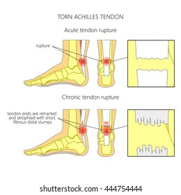
The peroneal tendons run down together behind the outer side of the ankle and then split before attaching to different parts of the foot. Hi, for the last week or so, i get this sudden sharp and pulsating pain in my foot, the inner arch area (left feet).i looked at a foot muscle diagram to identify the area and it looks like the abductor hallucis area where the pain is located. Ligaments are strong connective tissue composed of fibrous tissues. A foot tendon tear happens when one of the tendons in the foot is damaged from sudden injury or overuse. A foot or ankle injury can cause tendonitis.
Tendons Foot High Res Stock Images Shutterstock from image.shutterstock.com Flat feet or high arches can cause certain muscles to be out of balance. Familiarity with the normal anatomy of the plantar tendons and its appearance at magnetic resonance (mr) imaging and ultrasonography (us) is essential for recognizing plantar tendon disorders. Lateral view of foot & ankle. A tendon is band of tissue made up of many fibers. Pain and tenderness are concentrated on the top, bottom or the sides of your foot near the arch. The two main extensor foot tendons are the extensor hallucis longus and the extensor digitorum longus. Foot tendinitis is a very common cause of pain due to irritation or inflammation in tendons of the foot after overuse or injury. There are a whole range of structures e.g.
Tendinitis is treated with stretching and relaxing activities and exercises to give quality to the calf and foot muscles.
One peroneal tendon attaches to the outer part of the midfoot, while the other tendon runs under the foot and attaches near the inside of the arch. Bones, muscles, tendons and nerves which will each give slightly different foot pain symptoms. Swelling and bruising will occur at the site of injury. A tendon is a band of tissue that connects a muscle to a bone. A torn ligament of tendon in the foot is an injury that can limit daily activity. The muscles at the top of the foot fan out to supply the individual toes. Foot tendinitis is a very common cause of pain due to irritation or inflammation in tendons of the foot after overuse or injury. Ligaments are strong connective tissue composed of fibrous tissues. In the foot, there are two sesamoid bones located directly underneath the first metatarsal head, embedded in the medial (tibial) side and lateral (fibular) aspect of the flexor hallucis brevis tendon. The human foot combines mechanical complexity and structural strength. It connects muscle to bone. If you would like to learn all the parts of the foot structure, you have come to the right place. The two peroneal tendons in the foot run side by side behind the outer ankle bone.
There are a whole range of structures eg tendon diagram. Hi, for the last week or so, i get this sudden sharp and pulsating pain in my foot, the inner arch area (left feet).i looked at a foot muscle diagram to identify the area and it looks like the abductor hallucis area where the pain is located.